


Credit: Diana Sousa, Michael Hall


TEM images of the mesoporous silica nanoparticles

Credit: Catarina Martins

Breast cancer spheroid. Credit: Jhenifer Oliveira


Credit: Jhenifer Oliveira

Credit: L. Di Filippo, B. Mendes

Credit: L. Di Filippo

Hydrogels for urogenital infections. Credit: B. Mendes, J. Conniot


PhotoThermal performance of gold nanorods. Credit: R. Prasad, B. Mendes

Hyaluronic acid hydrogels for pancreatic cancer. Credit: J. Conniot, B. Mendes, J. Ravasco

lung metastasis from melanoma

Colon cancer cells full of gold nanoantennas (green)

Nanovesicles for monitoring metastasis

Credit: Bárbara Mendes, João Conniot, Diana Sousa

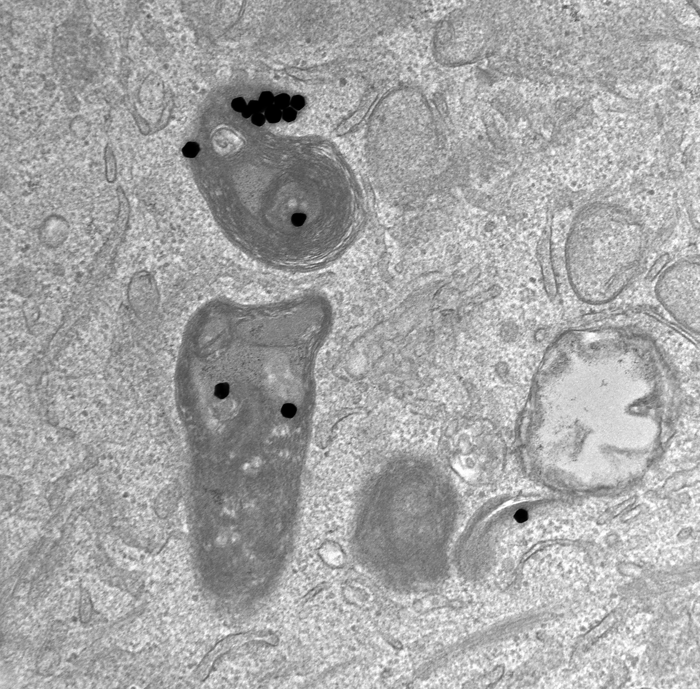
Electron microscopy image of cancer cell

Credit: Marta Santos


Photothermal images of NIR irradiated gold nanostars. Credit: Alessia Privitera


Credit: Bárbara Mendes, João Conniot

Credit: Bárbara Mendes, João Conniot

Credit: Jhenifer Oliveira, Bárbara Mendes

In vivo X-ray computed tomography 3D scans


Mice induced with Glioblastome multiforme cells

Gold Nanoparticles-miRs (red) invade cells (green) and relocate to the nucleus (blue) in 3D human cell culture model


Colorectal tumor (in red) on zebrafish larvae

Histology image of mice brain implanted with therapeutic hydrogels for glioblastoma

Confocal microscopy image of zebrafish larvae bearing colon cancer tumors (in red)

High-resolution SEM image of RNA triple-helix microstructures

siRNA nanoparticles (green) scavenged by tumour-associated macrophages expressing VEGF (red), a protein that helps tumours form new blood vessels to get nutrients

Dual-colour hydrogel scaffolds made of RNA-triple-helix nanoconjugates pre-incubated with complementary miR targets

Breast cancer cells (in red) internalized by miRNA-gold nanoparticles (in green).

Cryosection of dendrimer–dextran adhesive hydrogel depicting adhesive morphology. Red spots represent the triple-helix nanoparticles.

Colorectal cancer cells transfected with siRNA gold nanoparticles

High-resolution SEM image of RNA triple-helix microstructures

Tumor tissue impregnated with smart gold nanorods (in green) and gold nanospheres (in red).

Thermal imaging of mice implanted with a gel doped with smart nanorods that can heat up when illuminated in NIR

TEM image of +/- 20 nm gold nanoparticles

ATR-FTIR image of nanobeacons accumulated on lung tissue.

Gold nanobeacons (in red) able to image and detect a gastric tumor in vivo

Intracellular uptake of PAMAM dendrimers on cancer cells

Implantable hydrogel embedded dark-gold nanoswitch as a theranostic probe to sense and overcome cancer multidrug resistance.

Dark-field imaging of cells internalized with PEG-gold nanoparticles.